Tag Archives: Rehabilitation

Low Carb VS High Carb Dieting

High Carb vs Low Carb Diets… Interesting Case Study

I know many of you continue to feel as though the way to lose weight and/or maintain lean muscle mass is to minimize your carbohydrate intake.  Take a look at this article, written by Dr. S. Nadolsky, who is not only a physician but an avid body builder as well.  Dr. Nadolsky states, “…If you’re a healthy exerciser whose blood sugar levels are normal and you’ve been eating low carb for a while, I recommend trying a higher carb diet. You might be surprised at the results…”.  He definitely sheds some light on how and why a diet higher in carbohydrates may benefit us.

Carbohydrate confessions:  Stories (and data) from a low carb convert – By Dr. S. Nadolsky


Conclusions:

  1. Mindful eating makes the difference
  2. Weighing, measuring and tracking are important (I recommend this to anyone trying to make any type of physical change by changing their diet)
  3. Note that most lay people who are relatively inactive and somewhat overweight, should still stick with lower carbohydrate diets because it is much easier to get blood sugar and blood pressure controlled on a low carb diet
  4. while low carb diets have their place, I no longer think they’re necessarily the right choice, or the only choice, for everyone

Take home points:

  • Do not overly restrict; do not over-think it; do not waste time with detailed “carb math.”
  • Enjoy a wide variety of minimally processed, whole and fresh foods.
  • Observe how you look, feel, and perform.
  • Decide what to do based on the data you collect about yourself, not on what you think you “should” do.
  • The only “rules” come from your body and your experience. Do not follow a dietary prescription for anyone else’s body.

And above all, for most active people, carbs are your friend!

THE Stretch for Those That Sit

THE Stretch for Those That Sit

Target all of your sitting muscles at once with this smart move.

Perform this stretch as a dynamic warm-up before your regular cardio and/or strength sessions. Or you can perform it as a cool-down, but rather than walking through the movement, make it static and hold the final position (with ankle over knee) for a minimum of 30 seconds. 

Follow these step-by-step instructions:

       
(1) Stand with your feet together, arms at your sides.  

   
(2) Keep your back tall, shoulders down and chest open as you push butt behind you and bend knees slightly, going into a mini squat. Lift left foot a few inches off floor (as shown).  

(3) Hinge forward from hips to grab left knee with left hand and ankle with right hand. Slowly stand back up, bringing knee up with you, pulling it toward midline of chest, until your spine is fully extended (as shown).                                

   
(4) Then do another mini squat, pushing hips behind you, and place left ankle over right knee, with left knee out to side (as shown). 

(5) Extend arms at shoulder level in front of you and go deeper into your squat, pushing hips back, hinging forward from hips and reaching arms diagonally toward floor (as shown). Stand up, lunge forward with left leg, and then repeat stretch on the right. Continue alternating sides for 12 to 16 reps total (6 to 8 each leg).


  • To get more of a lat stretch: Reach arms away from the hip you’re stretching (i.e., if right ankle is over left knee, reach arms to left and push hips to right).
  • To make it easier: Use a wall for balance and/or do not go quite as deep into each squat.
  • To make it more difficult: Slowly work your way into a deeper squat. Or when you bring knee into chest, go up onto your tiptoes, which will target your calves and better activate your core and other stabilizing muscles.
Thanks to Linsdey Emery at equinox dk-apotek.com.com and Master Trainer Josh Stoltz

Flank pain

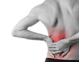
A patient of mine recently complained of flank pain, which was located in the right side near the lower back.  He trains in the martial arts and is often physical throughout his work week.  Martial arts training often encompasses movements which require the core muscles to be activated (kicking, punching, any type of grappling, weapons work, etc.).   My initial thoughts on a diagnosis were muscle/spinal-related however one must be very aware of other causes of flank pain.

flank pain

What Is Flank Pain?

Flank pain refers to pain or discomfort in your upper abdomen or back. It is located below the ribs and above the pelvis and on the side. Flank pain basically refers to pain in your side and back. Usually, the pain is worse on one side of your body.

Most people experience flank pain at least once in their life, and it is usually short lived. However, constant or severe flank pain may be caused by a serious medical condition, such as an infection in the urinary tract or kidneys, or dehydration. If severe flank pain occurs suddenly it could be from kidney stones. If it is chronic then it could be from several other causes.

Flank pain is often the sign of kidney problems, but it can also point to other medical conditions if it occurs along with other symptoms. If you experience chronic flank pain, it is important to talk to your treating health care practitioner and go over your symptoms.

What Causes Flank Pain?

Various conditions, ranging from serious to harmless, can result in flank pain.

Some common causes include:

  • arthritis (especially arthritis that affects the spine)
  • infection in the spine
  • spinal fracture
  • disk disease
  • spinal subluxation (causing nerve compression in the back)
  • muscle spasm (possibly due to spinal subluxation)
  • kidney infection
  • kidney stones
  • kidney abscess
  • shingles
  • dehydration
  • conditions in the chest where pain is referred to the flank
  • pneumonia
  • pancreatitis and other conditions affecting organs in your abdomen
  • inflammatory illnesses of the bowel such as Crohn’s disease
  • occasionally a heart attack can cause pain in the flank

What Symptoms May be Associated with Flank Pain?

Flank pain may be achy, cramp-like, or colicky—meaning it comes and goes in waves. Typically, kidney stones cause pain that is colicky and extreme. In the flank pain from kidney stones the patient has trouble lying in one position comfortably.

Flank pain may be accompanied by other symptoms, such as:

  • rash
  • fever
  • dizziness
  • nausea
  • constipation
  • diarrhea
  • blood in the urine

You should call your doctor right away if you experience the following symptoms along with your flank pain:

  • chills
  • fever
  • vomiting
  • nausea
  • blood in the urine or stool
  • prolonged or excruciating pain

Dehydration is one possible cause of flank pain. Seek immediate medical care if you experience flank pain along with these symptoms of dehydration:

  • extreme thirst
  • lack of sweat output
  • dizziness
  • fast pulse
  • dry, sticky mouth
  • headaches
  • constipation
  • decreased urine output

Diagnosing the Cause of Flank Pain

During your appointment, your doctor will try to identify the condition causing your flank pain. Be prepared to answer questions about:

  • when the pain began
  • what kind of pain you are experiencing
  • what other symptoms you have
  • how often you experience the pain
  • if the pain is pain sudden and passing, or constant
  • if you have had a recent a decrease in output of urine or intake of fluids
  • Whether there is pain in other parts of your body

Your doctor may also use imaging scans and blood tests to diagnose your flank pain. Imaging scans—such as an ultrasound or X-ray—allow your doctor to look deep within your body. These scans can reveal problems in the organs, tissues, muscles, and bones. A scan is particularly important in the evaluation of kidney stones to look for obstruction. Your doctor may inject a contrast dye into your vein before the scan, in order to better view any obstructions within your veins and organs.

Other tests that may be performed are:

  • CT (computed tomography) scan of your abdomen
  • cystoscopy—a minor procedure that uses a small scope to examine your bladder
  • urinalysis—a simple urine test
  • urine culture—a test where a urine sample is checked in a laboratory for bacteria

Treating Flank Pain

Rest is the primary treatment for any form of flank pain. Minor flank pain can typically be treated with a combination of rest and physical therapy. Your treating practitioner may also recommend specific exercises you can do for quick relief from muscle spasms.

For pain caused by inflammation—such as with arthritis, a subluxated spine (nerve compression) and infections—the treatment will depend upon the condition apotheke-zag.de/. If the condition is determined to be from arthritis in your spine you could benefit from physical therapy and an exercise program. Treatment for any type of spinal subluxation (which may cause nerve compression) can be treated by your chiropractor.  Your physician may prescribe an anti-inflammatory medication.   Your physician may prescribe you anti-inflammatory medication. Depending on your examination results, it may also be necessary for you to have a surgical evaluation. If there is an infection you may require antibiotics. Often infections in the spine are very serious and require antibiotics and even hospitalization.

Both kidney infections and kidney stones may require hospitalization. If you have a kidney infection, you will be given antibiotics to rid you of the infection. In some cases, you may receive these antibiotics intravenously. If you have kidney stones, you will need to drink lots of fluids to encourage the passing of the kidney stone and you may be prescribed pain medications. In most cases, kidney stones do not require surgery.

If the kidney stone does not pass then lithotripsy (breaking up stones with high frequency sound waves) may be used to break up the stones within the kidney. Once the stones are broken down by lithotripsy then they can be passed through the ureters (the tubes that carry urine from the kidney to the bladder). Other surgical techniques may also be used to remove the stone.

Always seek immediate medical attention when you develop sudden and intense flank pain.

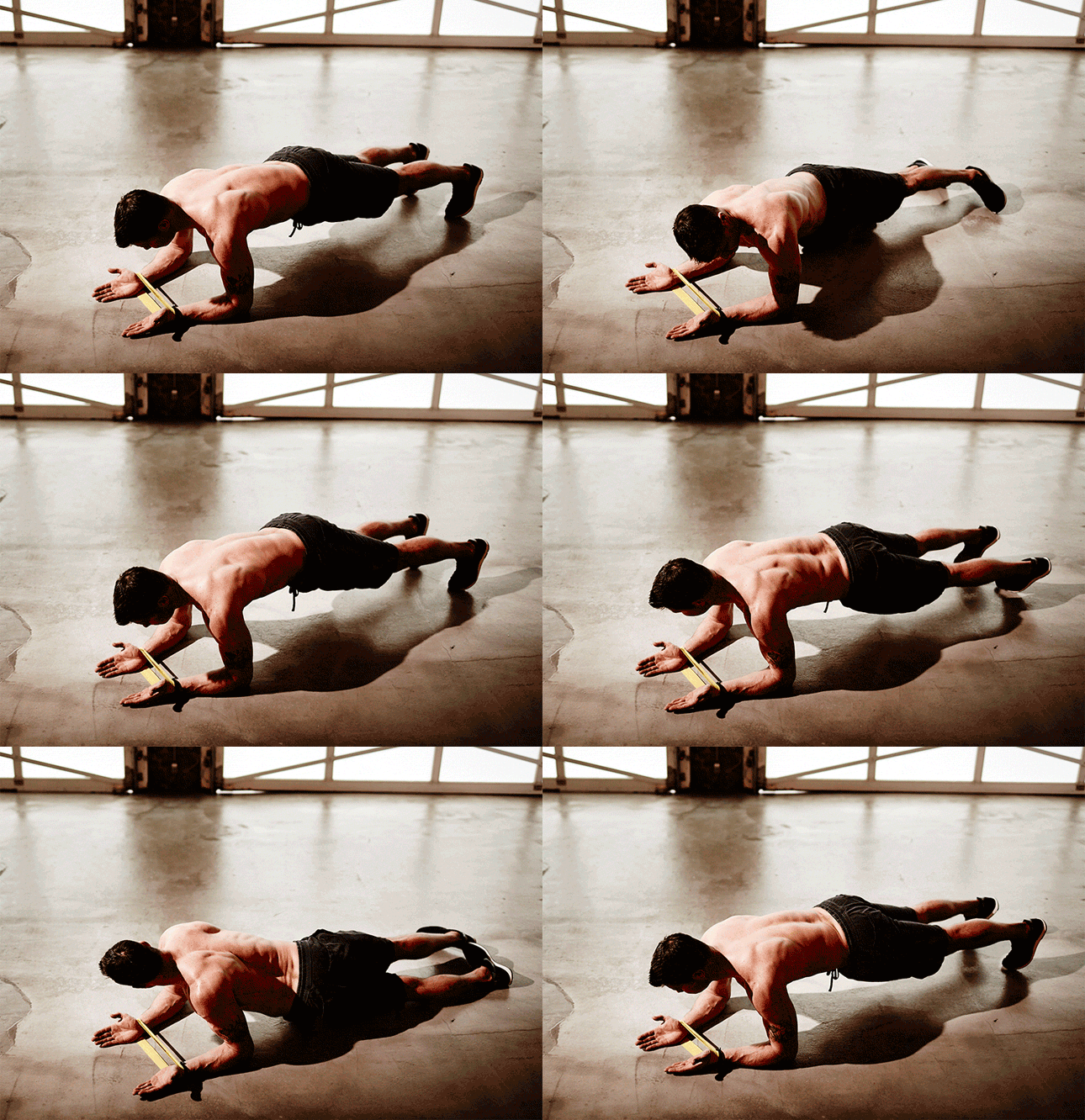
CORE EXERCISES – 3 Body-Changing Planks

CORE EXERCISES –  3 Body-Changing Planks

If you are looking to strengthen your core musculature (abdomen, lower back, pelvis, upper back/shoulders), take a look at these CORE exercises.  Please remember, when attempting new exercises, be very careful to activate the muscles that you are trying to work and remember to breathe correctly.  Core stabilization exercises activate many muscle groups at once and can be very challenging!

Acromioclavicular Joint (AC Joint) Injuries

AC Joint Injury

A good friend of mine recently injured his shoulder.  After assessing him, I realized that he had a Grade 1 AC joint injury.  After discussing his injury with him, as well as options for his rehabilitation, I decided to provide a little more information below, that you, the reader, could refer to at any time to help guide what may be an AC joint injury for you as well.

What is the AC joint?

The acromio-clavicular (AC) joint is the joint formed between the clavicle (collarbone) and the acromion (the tip of the shoulder blade which extends to the top of the shoulder). You can feel it, if you put your hand on top of your shoulder – it is the bony bump about 4cms from the edge of the shoulder.

The AC joint is a link between the arm and the trunk (your shoulder blade is closely connected to your rib cage by many different muscles) and is the only bony join between the shoulder blade and the rest of the body. It helps transmit load from the arm to the trunk in pushing, pulling, punching and resting on the arm.

AC joint

How the AC joint is injured?

The AC joint is a quite common sporting injury especially in contact sports. It is usually injured by a fall directly onto the shoulder, a fall onto the arm or a tackle.

The ligaments that bind the clavicle to the acromion are firstly stretched, then torn. Depending on the severity of the injury the clavicle can tear away from the acromion causing a noticeable lump to appear on top of the shoulder. The injury results in considerable pain, swelling and loss of shoulder movement. Depending on the severity of the injury, it will heal by itself or, if complicated, surgical intervention may be required.

Grading of an AC joint injury:

The most commonly used classification system recognises 6 severities of AC joint injury.

Grade I

A slight displacement of the joint. The acromioclavicular ligament may be stretched or partially torn. This is the most common type of injury to the AC Joint.  You will still have point-tenderness at the AC joint with palpation

grade 2 displacement AC jointGrade 2

A partial dislocation of the joint in which there may be dome displacement that may not be obvious during a physical examination. The acromioclavicular ligament is completely torn, while the coracoclavicular ligaments remain intact edmedicom.com.

grade 3 displacement AC joint.Grade 3

A complete separation of the joint. The acromioclavicular ligament, the coracoclavicular ligaments and the capsule surrounding the joint are torn. Usually, the displacement is obvious on clinical exam. Without any ligament support, the shoulder falls under the weight of the arm and the clavicle is pushed up, causing a bump on the shoulder

Grades I-III are the most common. Grades IV-VI are uncommon and are usually a result of a very high-energy injury such as ones that might occur in a motor vehicle accident.


Treatment for an AC joint injury

Initial treatment may consist of:

  • ICE (I place this modality first because icing is critical to maintaining minimal inflammation to allow the most effective environment for the body to heal)
  • Rest
  • Compression
  • Support (a sling may be worn)
  • Movement within the pain free range will help in maintaining mobility of the surrounding structures.
  • Taping may be beneficial to support the position of the joint.
  • Physical therapy can use ultrasound and interferential currents, for pain and inflammation.  Range of motion exercises within a pain free range will allow the ligament mobility as well.

As pain settles:

  • Load bearing exercises can be added to restore the normal function of the joint and surrounding muscles.
  • Massage and mobility exercises may be incorporated to ensure normal function is achieved.

In severe cases where the clavicle is completely torn away from the acromion the joint may remain painful and unstable and require surgical fixation.

Returning to sport following an AC joint injury:

Return to sport is possible when you have no localized tenderness, and full range of pain free movement has been achieved. On initial return to sport you may feel more comfortable to use taping or to have some padding over the AC joint. Your physical therapist can guide you on your return to sport and any precautions that need to be taken.

Examples of tasks you should be able to perform painfree are:

  • Landing against a wall sideways with your shoulder.
  • Landing against a wall onto an outstretched hand.
  • Throwing and catching a ball in awkward positions.
  • Completing one or more full contact training sessions.

Remember

  • Seek treatment at an early stage
  • ICE, ICE, ICE, ICE……and MORE ICE to decrease the inflammation!
  • Make sure that your diet is clean and healthy.  The old saying, “you are what you eat“, is absolutely true.  When you are rehabilitating from an injury, you must make sure that your body is being provided the necessary nutrients to heal.  It is imperative to understand that your body will NOT heal if you do not provide it with the necessary, and correct, nutrients!! (I will try to post more about nutrition, sport, recovery and physical rehabilitation in the future)
  • Ensure you physical therapist provides you with methods of self treatment and management.

If you have any questions regarding this information or your therapeutic management, please don’t hesitate to comment in an effort to create dialogue.


The information provided is for general information and does not substitute the advice and information your physical therapist will provide about your particular condition. While every effort has been made to ensure the information provided is correct and accurate, Dr. David Rick accepts no responsibility.

A PILOT STUDY DETERMINING THE INFLUENCE OF CERVICAL MANIPULATION ON SPINAL MOTION DURING GAIT IN PREVIOUSLY CONCUSSED INDIVIDUALS

A PILOT STUDY DETERMINING THE INFLUENCE OF CERVICAL MANIPULATION ON SPINAL MOTION DURING GAIT IN PREVIOUSLY CONCUSSED INDIVIDUALS – Copyright by Dr David Rick August 2005

For those in the world of academia, if you are interested in sports and concussion-related studies, please see the work I performed in 2005 which looked at chiropractic manipulation and spinal motion in previously concussed individuals.  The results of the study showed an increase in cervicothoracic motion (neck/upper back) after cervical manipulation.  Although the study only measured biomechanical factors (ie. neck/back motion), I would like to perform follow up studies on blood work which could include any type of hormonal stressors including cortisol, as well as neurotransmitters including serotonin.  Cortisol has been found to increase blood sugar, suppress the immune system, and decrease bone formation – all “bad” things.  Cortisol, when you are under any type of stress (physical, psychological, emotional, etc.), inhibits serotonin.  Serotonin has been thought to be a contributor of feelings of well-being and happiness in humans – “good” things. I would hypothesize that after spinal manipulation, there would be a decrease in cortisol and an increase in serotonin production in the body.  This means that there would be a decrease in the “bad” (cortisol) and an increase in the “good” (serotonin), which would assist the body in any type of recovery process including physical rehabilitation.

Please take a look at the following link where my close friend and colleague, Dr. John Minardi, briefly discusses how a spinal adjustment can affect the body in more than a biomechanical way.

How a spinal adjustment can increase mood and behaviour – Dr. John Minardi, April 2014

If you have any questions, please feel free to comment, I would be happy to answer!


Rapid Rehabilitation of a Hamstring Strain: A Case Study

Rapid Rehabilitation of a Hamstring Strain: A Case Study.

In 2005 I was treating athletes at the Ontario Soccer Association, where there was a high population of leg injuries, including numerous hamstring strains.  I had the opportunity of writing a Case Study which was published in The American Chiropractor on how to treat a hamstring strain.  If you take a quick look, I still use this methodology in treating my patients today, and I get wonderful results!

Please feel free to comment!


Advanced Healthcare Management Inc. – www.ahminc.ca

Please visit our site at www.ahminc.ca to explore how we can assist you if you were involved in a motor vehicle accident.

 Print


Please feel free to leave a comment!